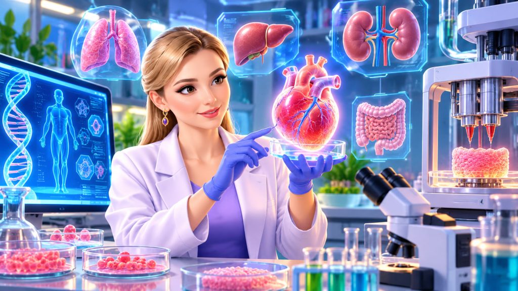
Imagine a future in which a damaged kidney can be replaced with a new one grown from a patient’s own cells. Instead of waiting years for a donor, doctors could potentially create living replacement tissue in a laboratory.
This remarkable possibility is driving some of the most exciting research in modern medicine.
Scientists are already growing miniature organs, printing living tissues, and developing biological replacements for damaged body parts.
The ultimate goal is to create functional human organs that can repair or replace diseased tissues without depending entirely on organ donors.
Although fully functional laboratory-grown hearts, kidneys, and livers are not yet available for routine transplantation, important breakthroughs are bringing this technology closer to reality.
What Are Lab-Grown Artificial Organs?
Artificial organs can be divided into two major categories.
Traditional artificial organs are mechanical or electronic devices designed to replace specific biological functions. Examples include artificial hearts and dialysis machines.
Laboratory-grown organs take a different approach.
They use living human cells to construct biological tissues capable of performing functions similar to those of natural organs.
This scientific field is called tissue engineering.
Researchers combine living cells, supportive materials, and carefully controlled growth conditions to encourage the formation of functional tissues.
The greatest challenge is recreating the extraordinary complexity of the human body.
A natural organ is not simply a collection of cells. It contains blood vessels, specialized structures, communication networks, and multiple cell types working together.
Step One: Obtaining the Right Cells
The first requirement is a reliable supply of living cells.
Scientists may collect cells directly from a patient’s body or use stem cells, which can develop into different types of specialized cells.
One particularly important innovation involves induced pluripotent stem cells.
These are mature body cells, such as skin or blood cells, that scientists reprogram into a flexible, stem-cell-like state.
Under appropriate laboratory conditions, they can develop into heart muscle cells, liver cells, nerve cells, and many other cell types.
Using cells derived from the patient could potentially reduce problems associated with transplant incompatibility.
However, researchers must carefully control cell development to prevent abnormal growth and ensure the final tissue functions correctly.
Step Two: Building a Framework for the Organ
Cells need a physical structure that guides their development.
Scientists often create a scaffold, a three-dimensional framework that supports cell attachment, growth, and organization.
These structures can be made from:
- Collagen and other natural biological materials
- Biodegradable synthetic polymers
- Hydrogels containing large amounts of water
- Processed tissue obtained from donor organs
One fascinating technique is called decellularization.
Researchers remove cells from a donated organ while preserving much of its natural structural framework.
The remaining structure can then be populated with new living cells.
This approach has been investigated for organs including hearts, lungs, livers, and kidneys.
Step Three: Printing Living Tissue in Three Dimensions
Another promising technique is 3D bioprinting.
Unlike conventional 3D printing, which uses plastic or metal, bioprinting deposits living cells and compatible biological materials in carefully arranged patterns.
Scientists often use a special printable material called bioink.
Bioink can contain living cells suspended in a soft gel that provides temporary support.
A bioprinter deposits this material layer by layer, gradually constructing a three-dimensional tissue.
Researchers have used these techniques to produce experimental skin, cartilage, and heart tissue structures.
However, printing a complete organ that can survive and function inside the body remains considerably more difficult.
Step Four: Helping the Organ Grow and Mature
Once cells have been arranged into a suitable structure, they must develop into functioning tissue.
Scientists frequently place their constructs inside a bioreactor.
This specialized laboratory system controls temperature, nutrients, oxygen levels, and other growth conditions.
Some bioreactors also provide mechanical stimulation.
For example, engineered heart muscle may benefit from electrical signals and physical stretching that imitate natural cardiac activity.
The objective is to encourage cells to organize, mature, and behave more like those found inside the human body.
Growing tissue is only the beginning; scientists must also teach it to function properly.
The Biggest Challenge: Creating Blood Vessels
One of the most difficult problems is vascularization, the formation of blood vessel networks.
Every large human organ needs a continuous supply of oxygen and nutrients.
Without adequate circulation, cells deep inside engineered tissue cannot survive.
Small pieces of laboratory-grown tissue can obtain nutrients through diffusion.
Larger structures require networks of blood vessels that connect successfully with the recipient’s circulation.
Scientists are investigating tiny printed channels, blood vessel-forming cells, and biological growth signals to address this challenge.
Creating reliable circulation is essential for producing transplantable hearts, kidneys, and livers.
Miniature Organs Already Exist in Laboratories
Researchers have achieved remarkable progress by developing organoids.
These are miniature, three-dimensional tissue structures grown from stem cells that reproduce selected features of real organs.
Scientists have created organoids resembling parts of the:
- Brain
- Intestine
- Liver
- Kidney
- Lung
- Retina
For example, brain organoids can help researchers investigate early neural development and certain neurological disorders.
Intestinal organoids allow scientists to study infections and test possible treatments.
Organoids are valuable research models, but they are not fully developed miniature replacement organs.
Most cannot independently perform all the functions required for transplantation.
Have Scientists Successfully Transplanted Lab-Grown Organs?
Some forms of tissue engineering have already reached patients.
Engineered skin has been used clinically to treat certain burns and difficult wounds.
In 2006, researchers led by Dr. Anthony Atala reported experimental bladder reconstruction using tissue-engineered structures produced from patients’ own cells.
Seven young patients with bladder abnormalities received engineered bladder tissue supported by biodegradable scaffolds.
The study demonstrated important possibilities for regenerative medicine, although the technique did not become routine clinical treatment.
Engineered cartilage and other relatively simple tissues have also been investigated in human clinical studies.
Complex organs remain a far greater challenge because they require multiple specialized cell types, precise architecture, and extensive circulation.
Expert Perspective: Why Function Matters More Than Appearance
Dr. Anthony Atala, director of the Wake Forest Institute for Regenerative Medicine, is one of the leading pioneers of tissue engineering.
His research has emphasized the importance of combining suitable cells, structural materials, and biological environments to create functional replacement tissues.
A major lesson from this work is that reproducing an organ’s appearance is not enough.
An engineered kidney must filter blood and regulate fluid balance. An engineered liver must perform essential metabolic functions. An engineered heart must contract reliably and sustain circulation.
A successful artificial organ must integrate into the body and perform its biological tasks safely over time.
Could Artificial Organs Eliminate Transplant Waiting Lists?
The long-term potential is enormous.
Thousands of patients worldwide depend on organ transplantation, while suitable donor organs remain limited.
Laboratory-grown replacements could eventually expand treatment options and reduce dependence on donated organs.
Using a patient’s own cells might also reduce immune rejection, the process through which the immune system attacks transplanted tissue.
However, cell-derived organs are not automatically immune-compatible, and producing them safely requires extensive testing.
Researchers must also solve problems involving manufacturing costs, tissue durability, infection control, and long-term performance.
Even with these challenges, regenerative medicine is steadily transforming the possibilities of human tissue repair.
Interesting Facts
- Japanese scientist Shinya Yamanaka’s team demonstrated cellular reprogramming in mouse cells in 2006 and human cells in 2007.
- The 2012 Nobel Prize in Physiology or Medicine recognized research showing that mature cells can be reprogrammed into a pluripotent state.
- Some laboratory-grown organoids can be maintained for months under suitable conditions.
- Researchers can use patient-derived organoids to investigate individual responses to certain medicines.
- Bioprinted tissues can contain several different cell types arranged in specific patterns.
- Engineered skin is among the most clinically developed applications of tissue engineering.
- Creating functional blood vessel networks remains a major obstacle to growing large transplantable organs.
- Laboratory-grown tissues may help scientists test drugs while reducing reliance on some animal experiments.
Glossary
- Tissue Engineering — The scientific process of creating or repairing biological tissues using living cells and supportive materials.
- Stem Cells — Cells capable of self-renewal and developing into specialized cell types.
- Induced Pluripotent Stem Cells — Mature cells reprogrammed into a state that allows them to develop into many different cell types.
- Scaffold — A three-dimensional supporting structure that guides the growth and organization of living cells.
- Decellularization — The removal of cells from biological tissue while preserving much of its structural framework.
- 3D Bioprinting — A technology that uses controlled printing methods to arrange living cells and biological materials into three-dimensional structures.
- Bioink — A printable biological material that may contain living cells suspended in a supportive gel.
- Bioreactor — A controlled laboratory system that supports the growth and maturation of biological tissues.
- Vascularization — The formation or development of blood vessels within living tissue.
- Organoids — Three-dimensional laboratory-grown cellular structures that reproduce selected characteristics of organs.
- Immune Rejection — An immune response that attacks transplanted cells or tissues recognized as foreign.