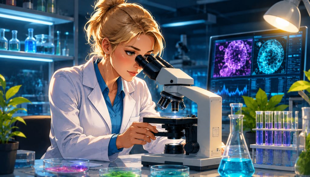
The microscope is one of humanity’s greatest scientific inventions. It transformed our understanding of life, disease, materials, and the Universe hidden beyond the limits of human vision. Before microscopes, people had no idea that bacteria, blood cells, or even the intricate structures inside plants and animals existed. Today, microscopes allow scientists to observe individual molecules, viruses, and even the arrangement of atoms.
The history of the microscope is a remarkable journey spanning more than two thousand years. From simple magnifying lenses used in ancient civilizations to today’s electron and cryo-electron microscopes capable of revealing structures measured in billionths of a meter, each technological breakthrough has opened new frontiers of discovery.
The Earliest Magnifying Lenses
Long before the invention of the microscope, people had discovered that certain transparent materials could magnify objects.
Archaeological evidence suggests that polished crystals and glass lenses were used in ancient civilizations, including:
- Ancient Egypt
- Mesopotamia
- Ancient Greece
- The Roman Empire
One famous example is the Nimrud Lens, a polished rock crystal dating back nearly 3,000 years. Although historians debate its exact purpose, it demonstrates that ancient people understood the basic optical properties of lenses.
These early magnifying devices were simple, but they laid the foundation for future optical instruments.
The Birth of the First Microscope
The first true microscopes appeared in the late 16th century.
Most historians credit Dutch spectacle makers Hans Janssen and Zacharias Janssen with developing one of the earliest compound microscopes around 1590.
Their instrument combined multiple lenses to achieve greater magnification than a single lens alone.
Although primitive by modern standards, this invention marked the beginning of microscopy.
The image quality remained poor because early lenses suffered from significant optical distortions.
Galileo’s Contribution
Shortly after the invention of the telescope, Galileo Galilei applied similar optical principles to create an improved microscope around 1609–1610.
His instrument, sometimes called the “occhiolino” (“little eye”), offered better magnification than many earlier designs.
Galileo demonstrated that scientific instruments could reveal hidden details of both the heavens and the microscopic world.
His work helped inspire further improvements in optical technology.
Antonie van Leeuwenhoek Changes Biology Forever
One of the greatest milestones in microscopy came during the 17th century.
Dutch scientist Antonie van Leeuwenhoek built remarkably powerful single-lens microscopes capable of magnifying objects more than 200 times.
Unlike many compound microscopes of his era, his carefully handcrafted lenses produced exceptionally sharp images.
Using these microscopes, he became the first person to observe:
- Bacteria
- Protozoa
- Sperm cells
- Red blood cells
- Microscopic algae
These discoveries fundamentally changed biology.
For the first time, scientists realized that an invisible world of living organisms surrounded them.
Robert Hooke and the Discovery of Cells
Around the same period, English scientist Robert Hooke published the landmark book Micrographia in 1665.
The beautifully illustrated work introduced readers to extraordinary microscopic images.
While examining thin slices of cork, Hooke observed tiny box-like structures.
He called them “cells” because they reminded him of small monastery rooms.
Although Hooke observed dead plant cells rather than living ones, his discovery introduced one of biology’s most important concepts.
Today, cell theory forms one of the fundamental principles of modern life science.
Better Lenses, Better Science
Throughout the 18th and 19th centuries, microscope technology advanced dramatically.
Scientists improved:
- Lens quality
- Glass manufacturing
- Optical alignment
- Mechanical precision
- Illumination systems
One major breakthrough was the development of achromatic lenses, which greatly reduced color distortion.
Later, German physicist Ernst Abbe established the mathematical foundations of modern microscope optics.
His work explained why microscopes have fundamental limits on optical resolution and guided the design of increasingly powerful instruments.
The Electron Microscope Revolution
Visible light eventually limits the maximum resolution of optical microscopes.
In the 1930s, scientists overcame this limitation by replacing light with beams of electrons.
Electron microscopes achieve far higher resolution because electrons have much shorter wavelengths than visible light.
Two major types are widely used today:
- Transmission Electron Microscope (TEM)
- Scanning Electron Microscope (SEM)
These instruments reveal structures thousands of times smaller than conventional light microscopes can resolve.
Scientists can now study:
- Viruses
- Cell organelles
- Nanomaterials
- Crystal structures
- Individual atoms under specialized conditions
Cryo-Electron Microscopy: A Modern Breakthrough
One of the most important recent advances is cryo-electron microscopy (cryo-EM).
Instead of staining or chemically fixing biological samples, researchers rapidly freeze them.
This preserves delicate molecular structures in a nearly natural state.
Cryo-EM has revolutionized structural biology by allowing scientists to visualize:
- Proteins
- Viruses
- Ribosomes
- Cellular machinery
At near-atomic resolution.
This technique has dramatically accelerated biomedical research, including drug discovery and vaccine development.
Modern Applications of Microscopes
Microscopes are now essential across countless scientific and industrial fields.
They are used in:
- Medicine
- Biology
- Chemistry
- Materials science
- Nanotechnology
- Electronics
- Forensic science
- Environmental research
Doctors use microscopes to diagnose diseases.
Engineers inspect semiconductor chips.
Geologists analyze minerals.
Biologists study living cells.
Without microscopy, many modern scientific disciplines would be impossible.
Expert Perspective
Physicist Professor Stefan Hell, recipient of the 2014 Nobel Prize in Chemistry, revolutionized microscopy through the development of super-resolution fluorescence microscopy, overcoming what had long been considered the fundamental diffraction limit of light microscopy. His work demonstrated that scientific creativity can push beyond seemingly impossible physical limits, allowing researchers to observe biological structures at unprecedented levels of detail.
Similarly, the 2017 Nobel Prize in Chemistry recognized Jacques Dubochet, Joachim Frank, and Richard Henderson for developing cryo-electron microscopy, a technique that transformed structural biology by enabling scientists to determine the three-dimensional structures of complex biomolecules with extraordinary precision.
A Window Into Invisible Worlds
The microscope has continually expanded humanity’s understanding of nature.
Each improvement has revealed new layers of complexity, from cells and bacteria to viruses, proteins, and atomic structures.
What began as a simple magnifying lens has evolved into one of the most powerful scientific instruments ever created.
As microscopy continues advancing through artificial intelligence, quantum technologies, and next-generation imaging systems, scientists will undoubtedly uncover even more of the invisible world that surrounds us.
The history of the microscope reminds us that some of the greatest discoveries are not found by traveling farther—but by learning to see smaller.
Interesting Facts
- The word microscope comes from the Greek words mikros (“small”) and skopein (“to look at”).
- Antonie van Leeuwenhoek discovered bacteria using handcrafted microscopes with a single tiny lens.
- Micrographia became one of the first scientific bestsellers thanks to its detailed microscope illustrations.
- Optical microscopes are limited by the wavelength of visible light, typically resolving details down to about 200 nanometers.
- Electron microscopes use beams of electrons instead of light, allowing much higher resolution.
- Cryo-electron microscopy has revolutionized drug development by revealing the structures of proteins and viruses.
- Modern super-resolution microscopes can observe structures previously thought impossible to see using visible light.
Glossary
- Microscope — An instrument used to magnify objects too small to be seen clearly with the naked eye.
- Compound Microscope — A microscope that uses multiple lenses to magnify an image.
- Achromatic Lens — A specially designed lens that minimizes color distortion caused by different wavelengths of light.
- Resolution — The ability of a microscope to distinguish two closely spaced objects as separate.
- Electron Microscope — A microscope that uses electrons instead of visible light to produce extremely high-resolution images.
- Cryo-Electron Microscopy (Cryo-EM) — An imaging technique that studies rapidly frozen biological samples at near-atomic resolution.
- Cell — The basic structural and functional unit of all living organisms.
- Super-Resolution Microscopy — Advanced optical microscopy techniques that overcome the traditional diffraction limit of visible light.